
Today, the peripherally inserted central catheter, PICC, is a widely used vascular acces device. Its use is spreading, thanks to a simple and fast insertion technique. This allows nursing teams to increase efficiency in the administration of long-term intravenous treatments.
But the use of PICC also carries some risks. Mostly:
- Thrombosis, when it is too short
- Arrhythmia, when it is too long
To minimise these risks and accurately check the position of the catheter tip, we can use four different methods:
- X-ray
- fluoroscopy
- echocardiography (transthoracic or transoesophageal)
- ECG catheter tip location.
X-ray
In the hospital it is an easily accessible resource, either in the radiology department or at the bedside. The catheter tip position is located and put in relation to anatomical references, which are not always clearly visible on the the X-ray.

In this method, the cavoatrial junction cannot be seen and is therefore subjective. This is shown in the study “Optimizing the patient positioning for PICC line tip determination” (Harako ME1, Nguyen TH, Cohen AJ), which proves that different professionals might interpret the catheter tip location in an X-ray quite differently. The length of the Superior Vena Cava can vary considerably from one patient to another: between 4.4 and 10 cm, and this is only one of the anatomical structures which vary from one patient to another.
Fluoroscopy
This method, which involves X-rays, is mainly used in the interventional radiology department. Performing a real-time X-ray gives a continuous, live image of the patient. The insertion of the catheter (navigation) can be visualised and the catheter tip can be located, to make sure it is in the right position.
Fluoroscopy is considered a sufficiently accurate method to position the central catheter. However, it is rather expensive: in terms of technology and devices needed. Moreover, it requires a X-ray proof room and trained personnel.
Its use is recommended only in special cases since it involves irradiation which should be avoided (INS).

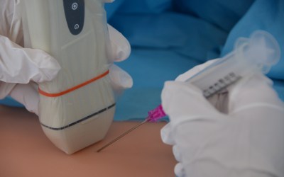
Transthoracic (TTE) and transoesophageal (TEE) echocardiography
Both types of echocardiography are safe techniques. TEE is the more accurate of the two.
A high level of expertise in ultrasound imaging is required to safely determine the position of the central catheter tip using echocardiography.
There is no standardisation of techniques. However, echocardiography is currently the most accurate method for locating the tip of a central line, surpassing the accuracy of fluoroscopy.
TEE is very unpleasant for the patient: it requires digestive fibroscopy to be able to place the US-probe intracavitary near the heart and can be applied thus to selected patients.
Compared to TEE, TTE is non-invasive. It depends more on the method used and on the practitioner. In terms of cost, an ultrasound machine is expensive, even more so with an echocardiography probe.

ECG system
Although it is amortised in a relatively short period of time, this system involves a significant initial cost which is expensive compared to, for example, a single X-ray. As with any ECG system, if it is located near other electrical devices, the image of the traces may contain artifacts. Using this method, we place the electrodes on the patients body in the same way as for any standard ECG monitoring.

The intracavitary electrode (the red or yellow clip is connected to the catheter) allows the catheter tip to be accurately located thanks to the interpretation of the P wave.

The location of the PICC catheter tip using an ECG system is the most appropriate technique for the following reasons:
• Easy to use: standard ECG monitor and use of few parameters
• Safe method: no additional risk in relation to the placement of the catheter itself
• Reliable and objective: P wave changes are observed, the technique is the same for everyone
• Quick and cheap: it is an intraprocedural method (the INS advises against a post-operative procedure) and the system is very affordable in the long run.
Bibliography
– Complicaciones del catéter según la posición de la punta Petersen et al, Am J Surg. 1999 Jul;178(1):38-41 Puel V et al, Cancer. 1993 Oct 1;72(7):2248-52 Eastridge et al, J Clin Oncol. 1995 Jan;13(1):233-8
– Cardiac Arrhythmias Resulting from a PICC: Two Cases and a Review of the Literature. Gapp J1, Krishnan M1, Ratnaraj F1, Schroell RP1, Moore D2. Cureus. 2017 Jun 3;9(6):e1308. doi: 10.7759/cureus.1308.
– http://www.ijccm.org/viewimage.asp?img=IJCCM_2010_14_4_180_76081_f6.jpg
– Aslamy et al, 1998 Sep;114(3):820-6
– Chu et al, Anesth Analg.2004 Apr;98(4):910-4 Wirsing et al, Chest. 2008 Sep;134(3):527-33
– Emerg Radiol.2004 Feb;10(4):186-9. Epub 2003 Dec 10
– http://www.medyrad.net/rayos-x-con-fluoroscopia
– Journal of Infusion Nursing: infusion therapy standards of practice. Gebhard RE, Szmuk P, Pivalizza EG, Melnikov V, Vogt C, Warters RD. The accuracy of electrocardiogram-controlled central line placement. Anesth Analg 2007; 104: 65-70.
– Pittiruti M, Bertollo D, Briglia E et al. Intracavitary ECG method for positioning the tip of central venous catheters: results of an Italian multicenter study. J Vasc Access 2012; 13(3): 375-65.
– Pittiruti M, La Greca A, Scoppettuolo G. The electrocardiographic method for positioning the tip of central venous catheters. J Vasc Access 2011; 12(4): 280-291.
– Rossetti F, Pittiruti M, Lamperti M, Graziano U, Celentano D, Capozzoli G. Intracavitary ECG method for positioning the tip of central venous access devices in pediatric patients: results of an Italian multicenter study. J Vasc Access 2015; 16 (2): 137-143.


0 Comments
Trackbacks/Pingbacks